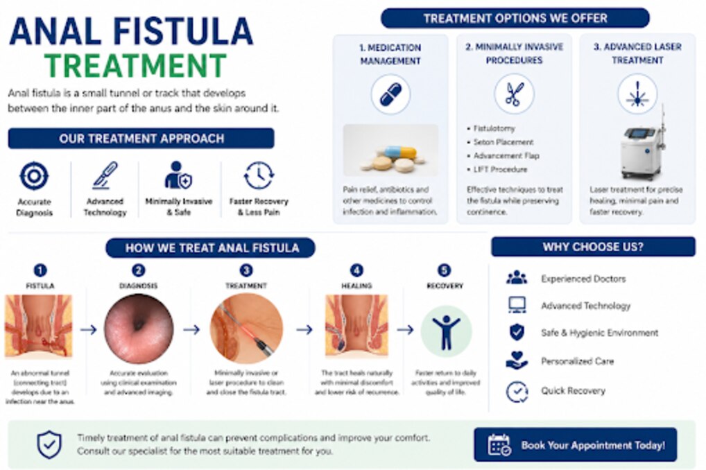
Choosing VAAFT depends on where the fistula runs, how much sphincter it crosses, whether branches or an abscess are present, and whether the internal opening can be treated securely. By the end, you will understand each operative step, the anatomy that affects eligibility, the alternatives, and the recovery trade-offs to discuss with a colorectal surgeon.
Key takeaways
- VAAFT enters through the external opening without cutting the entire tract open.
- MRI and examination help determine whether the tract suits a sphincter-preserving approach.
- High or branching fistulas may need another repair instead of VAAFT.
- Ask how recurrence and continence risks compare with fistulotomy for your anatomy.
What happens during a VAAFT procedure?
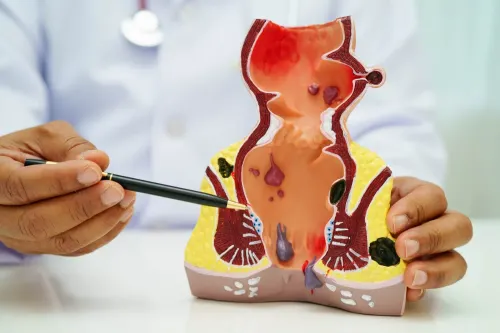
VAAFT is a sphincter-preserving endoscopic operation, not a camera-guided fistulotomy. After anaesthesia, you are positioned so the surgeon can examine the anal canal and reach the tract. The procedure starts at the external opening rather than by cutting the entire fistula open.
1. The surgeon gently widens the external opening and inserts the fistuloscope into the fistula tract. Irrigation clears blood, pus, and debris so the camera can show the tract lining.
2. The live video helps the surgeon follow the channel toward the internal opening inside the anal canal. The surgeon looks for side branches, dead ends, and abscess cavities instead of assuming that the most visible external opening represents the whole fistula.
3. Once the route is understood, the surgeon removes or destroys the inflamed tract lining using curettage, brushing, and electrocautery through the fistuloscope. This step reduces the tissue that keeps the channel open.
4. The surgeon treats the internal opening, usually by cleaning it and closing it with transanal sutures. This internal opening closure is essential: cleaning the outer tract alone can leave the source of faecal flow open and permit persistent drainage or recurrence.
The external opening is usually left to drain while the deeper repair heals, so a small opening or discharge immediately after surgery does not prove failure. Pain, bleeding, infection, urinary difficulty, and delayed healing remain possible even without a long open wound.
How the camera maps the tract and guides case selection
A camera can reveal whether the visible external opening leads to one continuous tract or a more complicated network. During video-assisted anal fistula treatment, irrigation opens the passage and clears pus or debris, allowing the fistuloscope to display the tract wall rather than relying only on an external probe.
1. The surgeon advances the fistuloscope from the external opening and follows the tract under direct vision. Changes in direction, narrowing, dead ends, and separate channels create the fistula tract mapping.
2. A second channel leaving the main passage indicates side branches. A wider, inflamed chamber containing pus suggests an abscess cavity; that finding can require drainage before definitive closure rather than simple treatment of the main tract.
3. The surgeon follows the tract toward the anal canal and identifies the internal opening as the point where it enters the bowel. A high, hidden, or multiple internal opening changes the repair plan because closing the wrong opening leaves the disease pathway active.
4. The video map is compared with examination under anaesthesia and, for complex, recurrent, or unclear disease, pelvic MRI. MRI can show extensions outside the field reached by the scope, so an apparently simple video view does not rule out deeper disease.
This map guides case selection: a clear, reachable internal opening and a tract that can be followed favour an endoscopic approach. Extensive branching, an undrained abscess, or anatomy the scope cannot define reduces certainty and may call for drainage or another repair strategy.
Which fistula anatomy suits VAAFT—and when does it not?
VAAFT is most reasonable in selected anal fistula cases where cutting through the sphincter could threaten continence. Examples include a high or transsphincteric fistula, a recurrent fistula, and disease in a patient with pre-existing sphincter weakness or previous continence problems. The internal opening must be identifiable, and the tract must be reachable with the fistuloscope.
| Finding | Likely direction | Reason |
|---|---|---|
| Simple, low tract with little sphincter involvement | Fistulotomy | Laying it open can provide reliable healing with limited muscle division |
| High or transsphincteric tract | VAAFT or another sphincter-sparing repair | Dividing more muscle increases continence risk |
| Active sepsis or undrained abscesses | Drainage, often with a seton | Sealing the tract before controlling infection can trap pus |
| Crohn’s disease, tuberculosis, malignancy, or radiation injury | Medical treatment, specialist management, or staged repair | Treating the underlying cause is essential for durable control |
| Unclear, branching, or recurrent anatomy | MRI, examination under anaesthesia, and tailored repair | A hidden extension or cavity can cause persistent drainage |
A branching tract does not automatically rule out VAAFT, but every extension and cavity must be assessed. Pelvic MRI becomes particularly important for recurrent or complex disease; the camera cannot replace preoperative mapping. If inflammation remains active, a draining seton may come first.
A fistula linked to Crohn’s disease may require medication before any definitive closure, while a large cavity, inaccessible internal opening, or tumour-related tract may call for another surgical strategy.
How VAAFT compares with fistulotomy and other repairs
The key choice is not “minimally invasive” versus “traditional.” It is sphincter preservation versus the certainty of laying a suitable tract open. Fistulotomy remains highly effective for a simple, low fistula crossing little sphincter muscle; cutting it open can provide more direct eradication than VAAFT.
| Option | Main trade-off | When it fits |
|---|---|---|
| Fistulotomy | Direct treatment, but divides involved sphincter muscle | Low fistulas with limited sphincter involvement |
| VAAFT | Preserves the visible sphincter ring, but closure can fail or miss branches | Selected high, transsphincteric, recurrent, or continence-sensitive cases |
| Seton placement | Drains and controls infection rather than definitively repairing the tract | Active abscess, ongoing sepsis, or staged treatment |
| LIFT procedure | Disconnects the tract between sphincter muscles without laying it open | Selected transsphincteric fistulas with an identifiable tract |
| Advancement flap repair | Covers the internal opening without dividing the sphincter | High or complex fistulas when flap tissue and healing conditions are suitable |
An active abscess changes the plan: drainage comes first. Seton placement can control discharge while inflammation settles, after which VAAFT, LIFT procedure, advancement flap repair, or another operation can be reconsidered.
VAAFT does not make continence risk zero. Infection, scarring, recurrent disease, sphincter manipulation, or an incorrect tract map can still affect function, and a second procedure may be needed. Reported studies are mostly observational with limited follow-up, so superiority over fistulotomy or other repairs is unproven.
At BariFit By Dr Parmeshwar, the useful comparison is anatomy-specific: ask how much sphincter the tract crosses, whether branches or abscess remain, and how previous operations and baseline continence change the risk.
Related service
Video-Assisted Anal Fistula Treatment (VAAFT) Video-Assisted Anal Fistula Treatment (VAAFT) Video-Assisted Anal Fistula Treatment (VAAFT) is an advanced minimally invasive procedure used for the treatment of anal fistula. View service → |
What recovery, recurrence, and continence risks should you expect?
VAAFT recovery is often easier than recovery after an open fistulotomy because the tract is not laid open through the sphincter. Expect soreness, light bleeding, or drainage from the external opening, and follow instructions for wound cleaning, pain relief, stool-softening, and activity.
- Fever, worsening pain, increasing swelling, or pus
- Difficulty passing urine
- Heavy bleeding or drainage that suddenly increases
A small external opening or continuing drainage does not prove early treatment failure. Healing is judged over follow-up visits by falling pain and discharge, closure of the external opening, and examination of the treated internal opening. Persistent symptoms may require examination under anaesthesia, pelvic MRI, or another procedure.
Published VAAFT healing rates vary because studies use different definitions, fistula types, follow-up periods, and combined treatments. Reviews commonly report primary healing of about 60% to 80% in the short to medium term, so fistula recurrence or persistent drainage remains a real possibility.
The principal advantage is avoiding deliberate division of the involved sphincter. That reduces, but does not eliminate, the risk to anal continence. Sphincter manipulation, infection, scarring, recurrent disease, or an incorrectly mapped tract can still affect control of gas or stool.
Ask how continence will be assessed before and after surgery, especially if you already have urgency, leakage, or prior sphincter injury.
Frequently asked questions
What happens during a VAAFT procedure?
After anaesthesia, the surgeon examines the anal canal, enters through the external opening, inspects the tract with a fistuloscope, cleans it, and closes the internal opening.
How does the camera guide VAAFT case selection?
The fistuloscope shows the tract’s internal opening, branches, and relationship to the sphincter, while examination and imaging help confirm whether VAAFT is appropriate.
Which anal fistulas suit VAAFT?
VAAFT is considered for selected fistulas where preserving sphincter muscle matters, including tracts that cross substantial sphincter muscle. Complex branching, active abscess, or anatomy the scope cannot safely access may require another treatment.
How does VAAFT compare with fistulotomy?
Fistulotomy lays open the tract and can work well for low fistulas, but it divides sphincter muscle. VAAFT aims to treat the tract while preserving that muscle, with a different recurrence and healing profile.
What recovery and continence risks should you expect?
Recovery often involves wound care, drainage, pain control, and follow-up until the tract closes. VAAFT limits planned sphincter cutting, but recurrence, persistent drainage, infection, and continence symptoms remain possible.